
If a carious lesion isn’t handled, demineralization can progress through numerous levels. Each stage indicates an exclusive level of severity in the demineralization system. The technique results in a cavitated lesion. Determining the stage of the lesion and whether a lesion is energetic or arrested can assist oral health experts in determining whether or not the tooth is a good candidate for dental sealant placement.
Fluoride is powerful in slowing or arresting the caries manner with the aid of contributing to the remineralization of tooth enamel. Early carious lesions, often called “white spot” lesions or incipient caries, are the earliest signs of tooth decay and can be arrested or managed before they progress to cavities. They appear as chalky white spots on the tooth surface, caused by demineralization of the enamel. Early detection and management can prevent further damage and the potential need for restorative treatments. Because fluoride is extensively handy (i.e., through network water fluoridation, fluoridated toothpaste, fluoride rinses, and fluoride varnish), it can take years for lesions to develop from one stage to the following, and many lesions can arrest (i.e., no longer progress to cavitation) or regress (i.e., remineralize).
Carious lesions may be categorized into two categories: non-cavitated and cavitated.
Non-Cavitated Carious Lesions
A non-cavitated carious lesion (also sometimes known as an early lesion, an incipient lesion, or a white spot lesion) is a demineralized lesion without evidence of cavitation. Lesions in the pits and fissures appear as a white/yellow/brown discoloration, are no longer regular with exogenous stain, and are confined to the pits and fissures inside the tooth floor. As the lesion progresses, the outer layer that is in touch with plaque and saliva is exposed to cycles of demineralization and remineralization, and the teeth regain some minerals (which include fluoride) and can grow to be less prone to additional breakdown.
Over time, the lesion will continue progressing underneath the tooth surface. At this level, the demineralization manner can be reversed or arrested through biochemical methods (e.g., fluoride use), mechanical methods (e.g., dental sealant placement), or both.
In its earliest tiers of improvement, a non-cavitated lesion may not be seen by the unaided eye. As the lesion progresses and turns into a cavity, it becomes visible to the unaided eye.
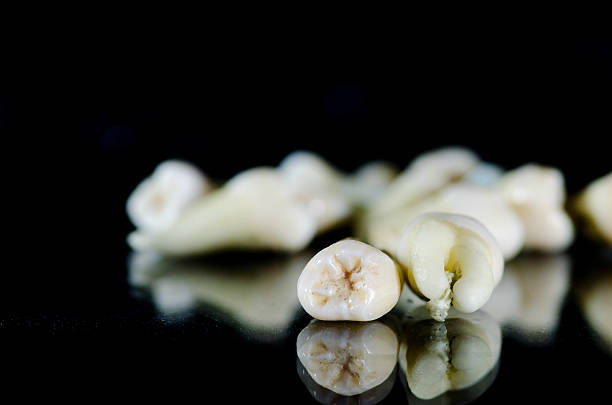
Cavitated Carious Lesions
Cavitated lesions (additionally called cavities) are lesions that have progressed to an advanced level and appear as a discontinuity or destruction in the floor due to a lack of tooth shape. Cavitation usually happens due to external forces that, in the end, result in the collapse of the outer enamel surface, which leads to a break or hollow in the teeth. The break inside the floor may be restrained to the teeth, or it could reveal the dentin.
By this factor, demineralization has usually stepped forward into the dentin, and microorganisms invade the dentin and develop into the pulp to purpose tissue contamination, also referred to as a dental abscess.
This stage of the disease normally calls for operative intervention to restore function and to assist in arresting the caries process within the tooth.
Active vs. Arrested Lesions
Carious lesions may be active or arrested. Active lesions showcase evidence of progression or trade over time, while arrested lesions do not. Thus, the most effective manner to decide with truth whether or not a lesion is active is to follow it through the years and look at its changes.
However, oral fitness professionals make clinical exams at a single point in time rather than following patients over the years. The following characteristics may additionally assist in figuring out whether or not a non-cavitated lesion is energetic:
- Active lesions tend to be whitish or yellowish in color and opaque (non-glossy). Inactive lesions may be whitish or yellowish and tend to be glossy.
- Active lesions feel difficult while the end of the explorer is moved lightly across their floor. Inactive lesions experience “tough” and “easy” while the top of the explorer is moved gently across their floor.
- Active lesions are positioned close to the gingival margin, and inactive lesions are located farther from the gingival margin, while the lesion is on a smooth floor.
When the lesion is on an occlusal (chewing) floor, it’s far more difficult to differentiate between an active and an inactive lesion. Such lesions are not easily seen by the unaided eye or detected with a gentle touch of an explorer. Drying the tooth with cotton rolls, gauze, or compressed air, while to be had, can also make it less difficult to look at the lesions. The oral fitness expert can check to see if the lesion is confined to the pits and fissures at the occlusal surface by looking for an underlying darkish shadow close to the grooves. The presence of a shadow suggests that the lesion is energetic and has extended into the teeth’s dentin.
Only energetic carious lesions require management. An energetic lesion does not continually develop to turn out to be a cavitated lesion with irreversible pulpal infection, and even when it does, the process is slow. Since active, non-cavitated lesions can be controlled with fluoride varnish and/or dental sealant utility, there’s no need to make quick and irreversible treatment selections. Early carious lesions are a significant concern for dental health, but with early detection and appropriate management, they can be successfully arrested or reversed, preventing further damage to the teeth and maintaining a healthy smile.